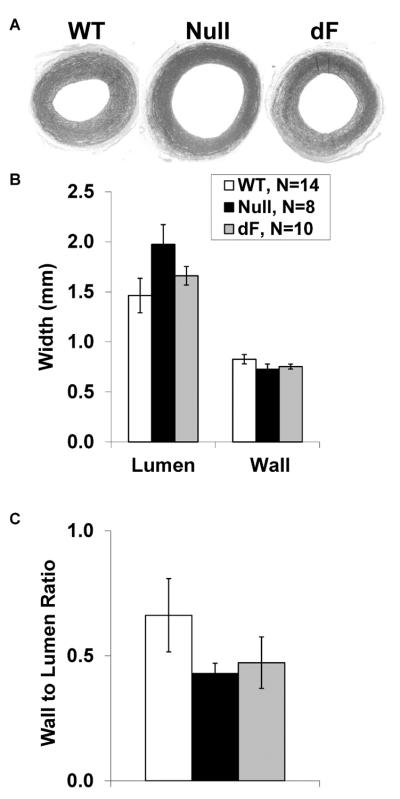
Figure 4.

Genotype-specific aortic morphology. Following dilation with sodium nitroprusside, aortic segments from WT (left, N=14), Null (middle, N=8) and dF piglets (right, N=10) were fixed and stained for morphometric analysis (A). While the aorta from CFTR-null piglets tended to have increased lumen diameters (B) and decreased wall to lumen ratios (C), these did not approach statistical significance.
