Fig. 6.
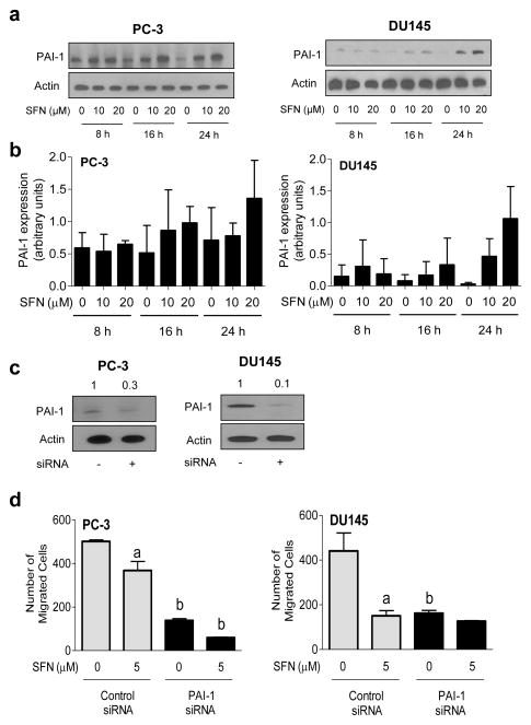
SFN treatment causes induction of PAI-1 protein in prostate cancer cells. a Western blotting for PAI-1 protein using lysates from PC-3 and DU145 cells after treatment with DMSO (control) or the indicated concentrations of SFN for specified time periods. b Densitometric quantitation of PAI-1 protein expression changes (arbitrary units) from western blots shown in panel a. Results shown are mean ± SD (n=2–3). Quantitation was normalized against actin protein band intensity for each individual experiment. c Western blotting for PAI-1 protein using lysates from PC-3 and DU145 cells transiently transfected with a control siRNA or a PAI-1-targeted siRNA. Blots were stripped and reprobed with anti-actin antibody. d Quantitation of cell migration in PC-3 and DU145 cells transfected with control siRNA or PAI-1-targeted siRNA after 24 h treatment with DMSO or 5 μM SFN. Results shown are mean ± SD (n=3). Significantly different (P < 0.05) compared with acorresponding DMSO-treated control and bbetween control siRNA transfected cells and PAI-1 siRNA transfected cells at each dose (0 or 5 μM SFN) by one-way ANOVA followed by Bonferroni’s multiple comparison test. Each experiment was repeated at least twice.

