Figure 1.
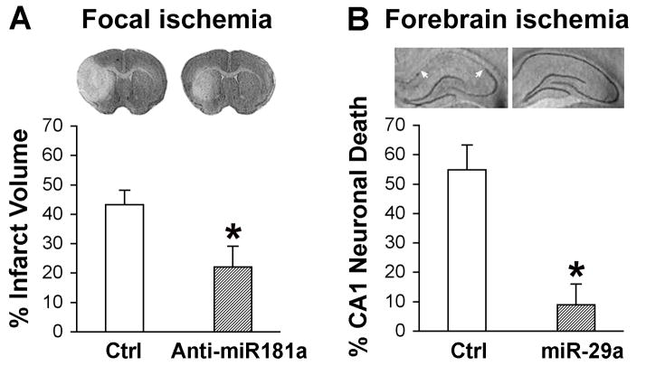
Effect of miR-181a and miR-29a on focal and global cerebral ischemia. A. Representative cresyl violet-stained coronal sections demonstrate a decreased infarct size in miR-181a down-regulated brain compared with brain treated with miR-181 mismatch (Ctrl) subjected to 1 h MCAO and 48 h reperfusion. The bar graph shows quantitation of infarct size (modified from Fig. 5 in [42]). B. Representative cresyl violet-stained coronal hippocampal sections demonstrate selective loss of CA1 neurons (between white arrows) at 6 days reperfusion after 10 min forebrain ischemia in control ischemic animals and protection with miR-29a overexpression. Quantitation of CA1 neuronal loss is shown in the bar graph (modified from Fig. 6 in [46]).

