
Figure 1. Background K+ channels in TG neurons.
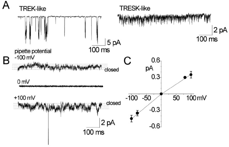
A. Cell-attached patches were formed on TG neurons (small to medium-sized cell bodies) and inward single-channel current recorded at pipette potential of +100 mV in bath solution containing 150 mM KCl. TREK- and TRESK-like channels are shown.
B. Small-conductance channels in cell-attached patches are shown at three pipette potentials (0 and ±100 mV). Dotted lines are drawn by eye to indicate open levels.
C. Amplitude of channel opening was measured at different potentials by drawing lines through open and closed levels, and plotted as a function of membrane potential.