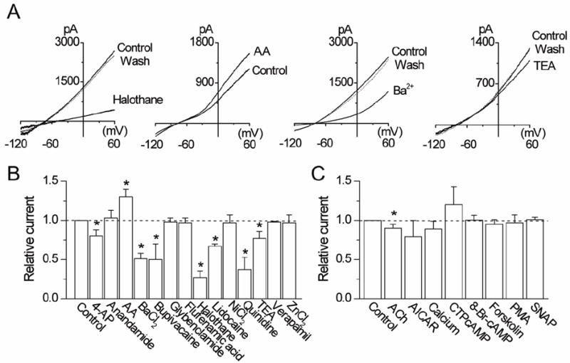
Figure 3. Effects of pharmacological agents on THIK-1 current.

A. Representative tracings show whole-cell currents before, during and after perfusion with halothane (2 mM), arachidonic acid (10 μM), Ba2+ (3 mM) and TEA (1 mM).
B and C. Relative changes in whole-cell currents measured at +60 mV are shown. Control is taken as 1.0. Each bar is the mean±SD of 5-10 determinations. Asterisk indicates a significant difference from the control value (p<0.05).
