Fig. 2.
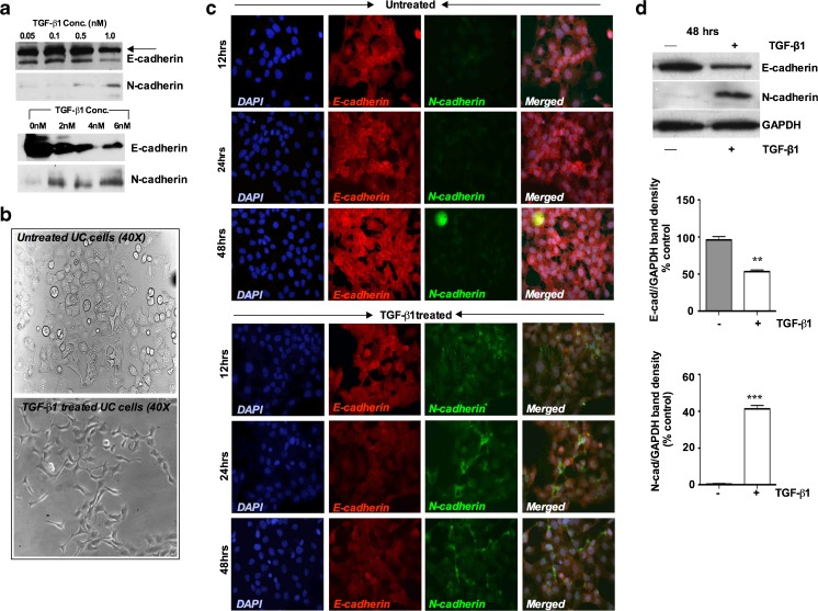
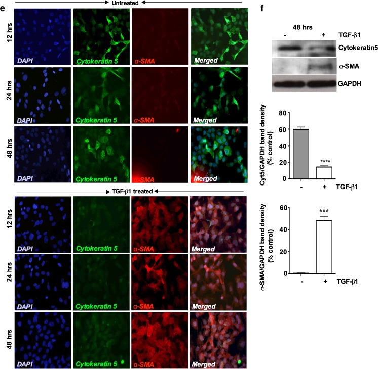
TGF-β1 treatment induces EMT-related genes in porcine bladder UC cells. a Western blot analysis UC cell lysates after different concentration of TGF-β1 treatment. Cultured UC cells were incubated with 0.05, 0.1, 0.5 and 1nM of TGF-β1 in the serum free medium and induction of EMT markers were assessed (Upper panel). Similar experiment was repeated in a higher concentration of TGF-β1 to assess the EMT induction. Cells were treated with 0nM, 2nM, 4nM and 6nM of TGF-β1 for up to 48 h. Western blot analysis showed no significance changes in E-cadherin and N-cadherin expression in lower TGF-β1 concentration. However, at 1nM and 2nM TGF-β1 concentration showed a decrease in E-cadherin expression whereas N-cadherin expression started to increase. b Morphology of the UC cells by phase-contrast microscopy after TGF-β1 stimulation. TGF-β1 treated cells showed a decrease in cell-cell contacts and adopted a more elongated and spindle fibroblasts like shape (Magnification X40). c Immunofluorescence staining showing switching of epithelial marker E-cadherin to mesenchymal marker N-cadherin in a time dependent manner Magnification × 40 (Scale Bar 2 μM). d Representative Western blots showing decreased expression of E-cadherin and increased expression of N-cadherin, in UC cells treated with TGF-β1 (6nM) for 48 h with quantitative densitometric analysis (p < 0.05). e Immunofluorescence staining showing loss of epithelial marker cytokeratin5 and gain of mesenchymal marker α-SMA after TGF-β1 treatment for 48 h (Scale bar 2 μM). f Representative Western blots showing decreased expression of cytokeratin5 and increased expression of α-SMA in UC cells after TGF-β1 treatment for 48 h, with quantitative densitometric analysis (p < 0.05)


