Fig. 3.
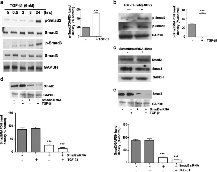
TGF-β1 stimulation induces phosphorylation of Smad2 and Smad3 in cultured porcine bladder primary UC cells: Porcine bladder UC cells were cultured as described in Materials and methods. TGF-β1 was added to the cultures and incubated for indicated times (0.5, 2, 8 and 24 h). a Western blots showing time (0, 0.5, 2, 8, 24 h) dependent activation of p-Smad2 and p-Smad3 after TGF-β1 treatment. b Activation of p-Smad2 and p-Smad3 48 h after TGF-β1 treatment compared to untreated (control) cells, with quantitative densitometric analysis (p < 0.01). Porcine bladder UC cells were transfected with Smad2 and Smad3 siRNA and incubated for 24 h at 37°C. TGF-β1 was added to the culture and further incubated for 24 h (total incubation time 48 h). Cells were lysed and expression of Smads was assessed by Western blot. c Smad2 and Smad3 showing unchanged expression of respective Smads in scrambled non-specific siRNA treatment. d and e siRNA treatment resulted in about 65 % reduction in the band density in respective proteins assessed 24 h after the start of transfection, with quantitative densitometric analysis (p < 0.01)

