
Fig. 2.
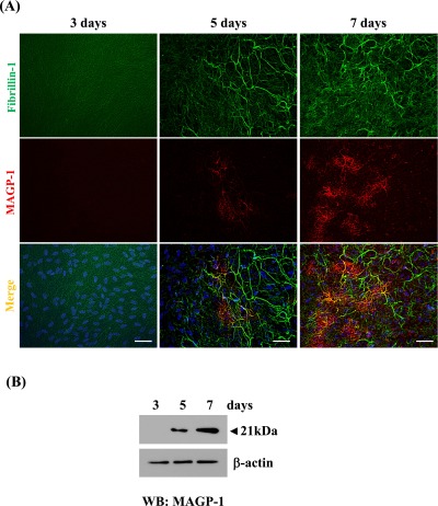
Immunolocalization and deposition of MAGP-1 to oxytalan fibers. (A) Double immunofluorescence for fibrillin-1 (upper lane) and MAGP-1 (middle lane) in cultures of human non-pigmented ciliary epithelial cells. Human non-pigmented ciliary epithelial cells were cultured, then simultaneously labeled for fibrillin-1 (green) (upper panels), MAGP-1 (red) (middle panels), and superimposition of both labels (lower panels) on days 3, 5 and 7. DAPI was used for nuclear staining (blue). Bar=20 µm. (B) Western blot analysis: Total cell/matrix proteins were extracted and 5 µg was subjected to Western blot analysis, as described in “Materials and Methods”. A 21-kDa band corresponding to MAGP-1 was detected on day 5 and the level of intensity increased up to day 7.