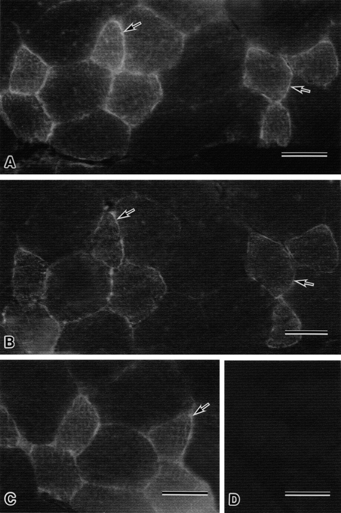
Fig. 3.

Immunohistochemical stainings with anti-AQP7, AQP4 and AQP9 antibodies of serial sections of muscle from wild mouse (A, B, C), respectively, and immunocontrol staining of wild mouse muscle (D). The immunostaining sample with anti-AQP7 antibody from wild muscle contains the myofibers reacted with anti-AQP7 antibody at their cell surface (arrows in A) and the myofibers without immunoreactivity by anti-AQP7 antibody (A). The serial muscle section immunostained with anti-AQP4 antibody (B) demonstrates that the fairly well stained myofibers at their cell surface with anti-AQP7 antibody observed in A (arrows) are also obviously immunoreactive with anti-AQP4 antibody (arrows in B), and therefore AQP7 positive myofibers are type 2 fibers. The serial muscle section with immunostaining by anti-AQP9 antibody reveals that the fairly well stained myofibers with this antibody observed in C (arrow) are also type 2 myofibers on the basis of evidence provided with figure B. The immunocontrol sample of wild muscle reveals no immunoreaction at the myofiber surface (D). Bar=50 µm (A–D).
