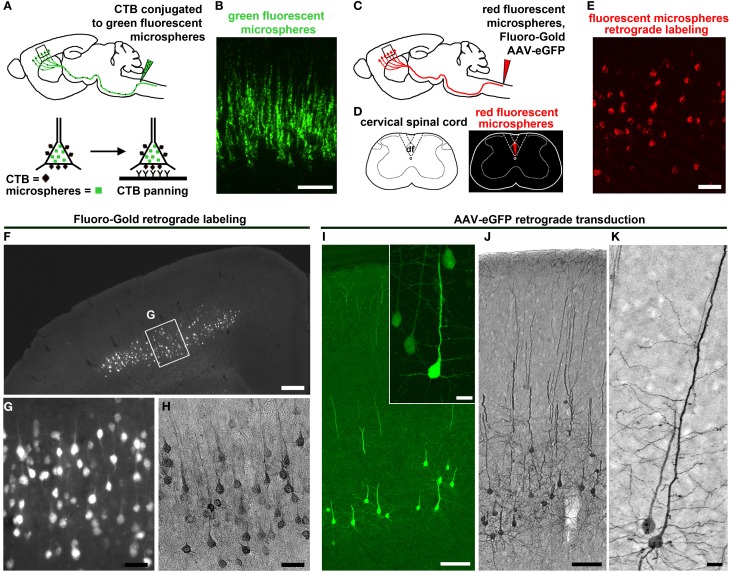
Figure 2.
CSMN retrograde labeling and transduction approaches. (A) Schematic drawing of CTB injection conjugated to green fluorescent microspheres into the pyramidal decussation of the spinal cord. Retrograde labeled CSMN containing CTB on their surface can be isolated by immunopanning. (B) Enlarge inset of the motor cortex enlarged to the right. Modified from Dugas et al. (2008). (C) Schematic drawing of retrograde labeling of CSMN by injection into the CST of the cervical spinal cord (C2). (D) Injection is restricted to the CST that lies within the dorsal funiculus (df). (E) Enlarged inset of the motor cortex showing CSMN retrogadely labeled with red fluorescent microspheres. (F) FG retrograde labeling. CSMN are visualized throughout the motor cortex by direct analysis on fluorescent microscope. (G) Enlarged inset of CSMN in the motor cortex. (H) CSMN cell body and proximal apical dendrite structures are enhanced by FG immunocytochemistry using DAB. (I) AAV-mediated retrograde transduction. CSMN are visualized throughout the motor cortex by eGFP immunocytochemistry using fluorescence microscopy. Large inset of a representative CSMN in the right corner. (J) CSMN cytoarchitectural structure is enhanced by eGFP immunocytochemistry using DAB. (K) large inset of a CSMN shows details of CSMN cell body, basal dendrites, apical dendrite and spines. Scale bars: (B) = 100 μm, (E) = 50 μm, (F) = 500 μ m, (G,H) = 50 μ m, (I) = 100 μ m (inset = 20 μ m), (J) = 100 μ m, (K) = 20 μ m.

