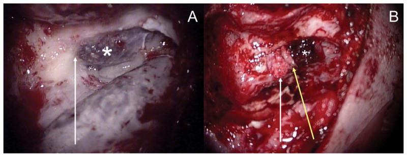
Figure 4.

A) Intra-operative view of the high right jugular bulb (asterisk) apposed to the inferior aspect of the otic capsule (arrow) prior to repair. B) After reduction of the high jugular bulb (asterisk), composite repair was performed by plugging of the PSCD with fascia followed by placement of bone pate (arrow) and a bone graft (yellow arrow).
