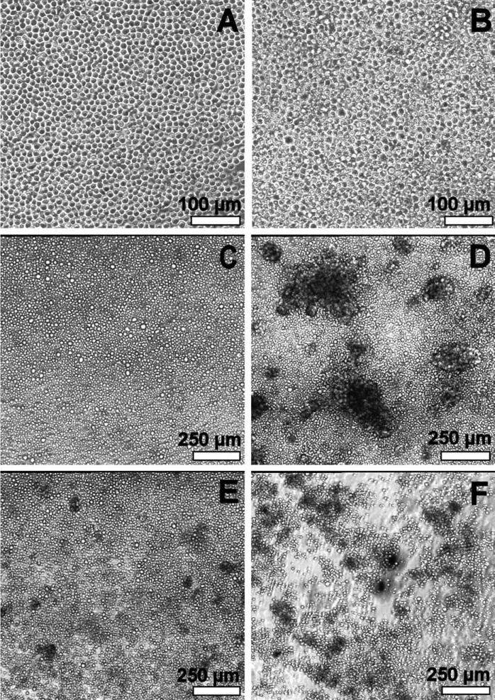
FIGURE 2.
Phase contrast microscopy. (A) Week 1: similar morphology on all surfaces. (B) Week 2: similar morphology on all surfaces. Some cells with larger diameter compared to week 1. (C–F) Day 17: polystyrene control, heparin, dermatan sulfate, and hyaluronan, respectively. Chitosan control and heparan sulfate surfaces exhibited cell morphology similar to the heparin surface.

