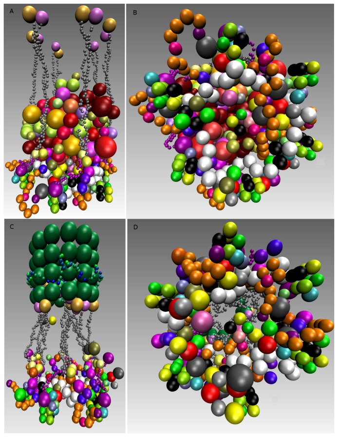
Figure 9.
Single rosette-like structure assembly in mitosis. (A) Shows a rosette-like structure where SACis active and no microtubule is present or attached; (B) Same as in Panel A, showing the rosette-shape in top view; (C) Shows a rosette-like structure attached to a microtubule and including the Ska complex, where the Spindle Assembly Checkpoint (SAC) switched off; (D) Same as Panel A, showing the rosette-shape in top view similar to Panel B, while SAC is inactive. The distance between two nucleosomes is 14 nm, and consequently, the diameter of the rosette-shape and microtubule is 25 nm. The rosette-structure and the Ska-complex are 60 nm apart. The attached, as well as the unattached kinetochore took about one hour to simulate.

