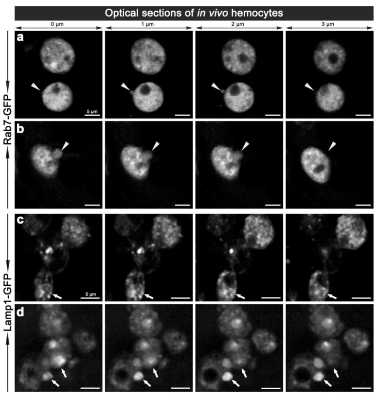
Figure 5.
Two-photon micrographs illustrating the in vivo morphology of adult hemocytes carrying CG-GAL4>Rab7-GFP (a, b) and CG-GAL4>Lamp1-GFP (c, d) markers. Each row shows consecutive optical 1 μm thin sections along the Z-axis with 0.5 μm steps in-between, which are representative of Movie 4 and 4a (a), and Movie 5 (b). Arrowheads (a, b) depict the vesicles protruding from the hemocyte surface. Arrows (c, d) depict small or enlarged Lamp1-GFP compartments. Scale bars = 5 μm.

