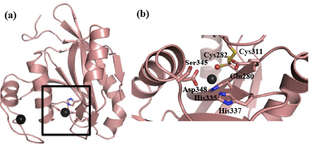
Figure 1.
The DUB domain of AMSH. (a) Ribbon diagram of the crystal structure of the catalytic domain of AMSH (PDB ID: 3RZU). The active site is highlighted by the black square. (b) An expanded view of the active-site residues of AMSH. The black spheres represent Zn2+, and the red sphere the active-site water molecule.

