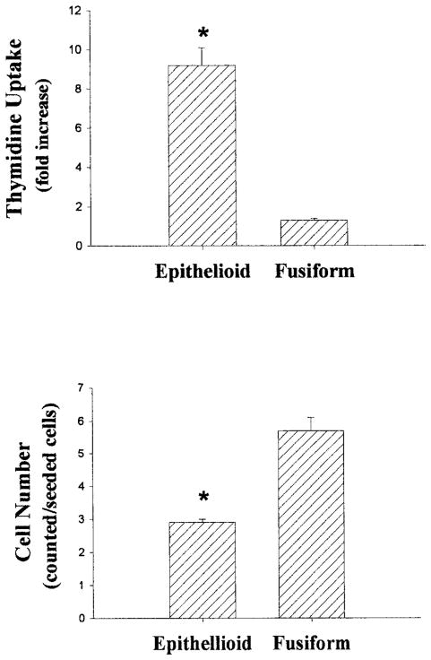
Figure 2.
Growth responses of fusiform and epithelioid SMC lines. For experiments shown in the upper panel, confluent SMCs were growth-arrested by incubation with serum-free medium for 48 hours. The cells were then incubated with or without 10% FCS for 24 hours, whereas [3H]thymidine uptake (expressed as fold increase) was determined by liquid scintillation counting. For experiments shown in the lower panel, SMCs were passaged into 24-well plates at an initial concentration of 5000 cells/cm2 in medium supplemented with 10% FCS. After 7 days, the cells were removed by trypsinization, and cell numbers (expressed as counted/seeded cells) were determined by using a hemocytometer. Data are expressed as mean±SE, n=3 per group, representative of 2 experiments. *P<0.05 vs fusiform SMCs.

