
Figure 5.
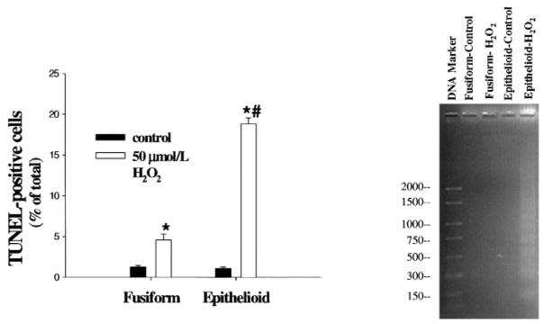
Effects of H2O2 on apoptosis in fusiform and epithelioid SMCs. After treatment with 0 (control) or 50 μmol/L H2O2 for 24 hours as described in the legend to Figure 2, the TUNEL assay was performed (left panel), or DNA fragments were extracted and subjected to agarose gel electrophoresis for assessment of laddering (right panel); the values to the left of the DNA marker refer to the numbers of base pairs. TUNEL-positive cells were quantified and expressed as the percentage of total cells in each cell line. Values are expressed as mean±SE, n=3, representative of 2 experiments. *P<0.05 vs corresponding control value. #P<0.05 vs fusiform SMC value at 50 μmol/L H2O2.