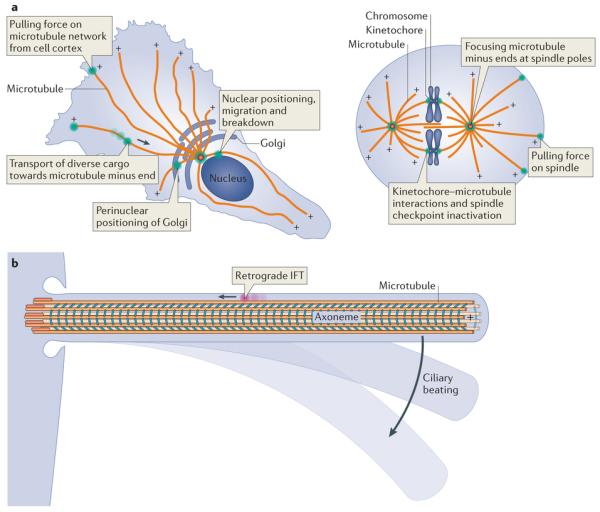
Figure 1. Sites of dynein action in the cell.

a | Example functions of cytoplasmic dynein (green) are shown in an interphase cell (left) and a dividing cell (right). The polarity of microtubules is indicated by plus signs. The arrow depicts the direction of dynein movement towards the microtubule minus end. Note that in some cell types and regions, such as the dendritic arbors of neurons, the microtubule network can have mixed polarity. b | Dynein functions in cilia. Intraflagellar transport (IFT) dynein (pink) performs retrograde IFT, whereas axonemal dyneins (cyan) power the beating of motile cilia.
