
Figure 2. Epidermal thickening and psoriasiform changes in CXCR4f/f control and K14-CXCR4KO mice following IL-23 injection.
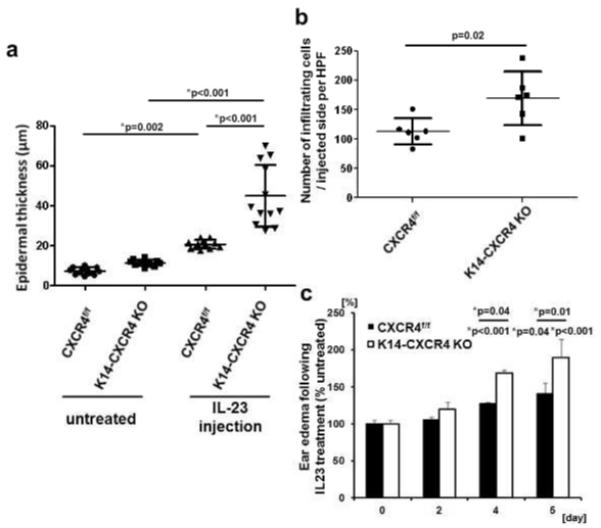
Ear sections from K14-CXCR4KO mice (n=3) and CXCR4f/f control mice (n=3) treated with IL-23 every other day for 5 days were H&E stained. The subjectively thickest epidermal regions were selected, and epidermal thicknesses were measured at four points in a blinded fashion for each mouse sample (A). Dermal infiltrating cells were counted on randomly selected sections of IL23 treated mice (n=3; B). Ear swelling was measured before injection (day 0, 2, 4) and harvesting ears (day 5; n=3). Representative data from one of two independent experiments with similar results are shown (A, B, C). *Indicates significance using Scheffé’s post hoc comparison test.