
Figure 4. Effects of CXCL12 on HaCaT cell proliferation, SOCS3 expression, and phosphorylation of STAT3.
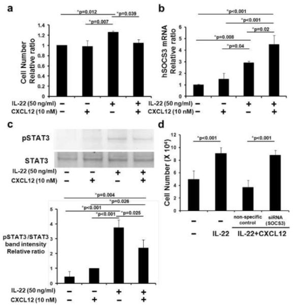
CXCR4-HaCaT cells were cultured for 1 day, then with human CXCL12 and/or human IL-22. After 2 more days (n=5 wells/condition), cells were counted. Summary of 3 independent experiments shown (A). CXCR4-HaCaT cells were stimulated with CXCL12 and/or IL-22 for 1 hr. SOCS3 mRNA expression was measured using real-time RT-PCR (n=3 wells/condition; 1 of 3 experiments shown, B). Phospho-STAT3 (pSTAT3) and total STAT3 were examined by Western blot (representative experiment shown, C). Analysis by densitometry (normalized using vehicle-treated cells) of 3 independent experiments is shown in the lower panel (C). CXCR4-HaCaT cells were transfected with SOCS3 siRNA and non-specific control siRNA, cultured with CXCL12 and IL-22 for 2 days, and counted (n=5; D). *Indicates significance using Scheffé’s post hoc comparison test.