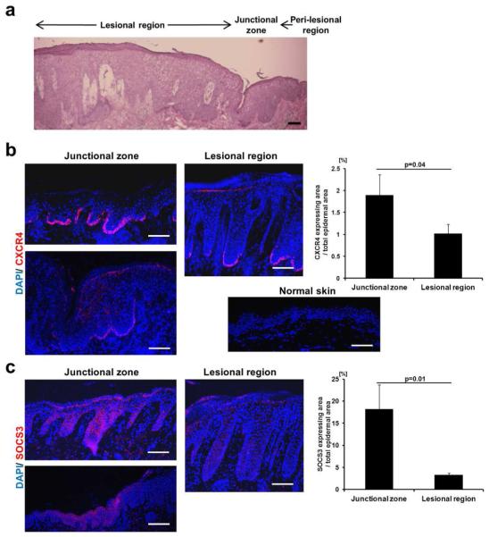
Figure 5. CXCR4 and SOCS3 expression in human psoriatic skin.
Three human psoriasis skin samples containing the junctional zone between lesional and perilesional regions were biopsied (see Supplementary Table S1.). An example of H&E stainings showing these regions is shown (Patient 1; A). CXCR4 (B) and SOCS3 (C) stainings of the junctional and lesional area were performed and representative images of similar findings are depicted (Patient 2; See Supplementary Figure S6.). A healthy human skin sample was stained using anti-CXCR4 Ab (B; lower-left panel). CXCR4- and SOCS3-positive areas of 3 psoriasis patients were quantified using imageJ software and used for calculation of ratio (%) of marker-positive area to epidermal junctional or lesional zone areas (Right panels of B and C). Scale bars, 100 μm.

