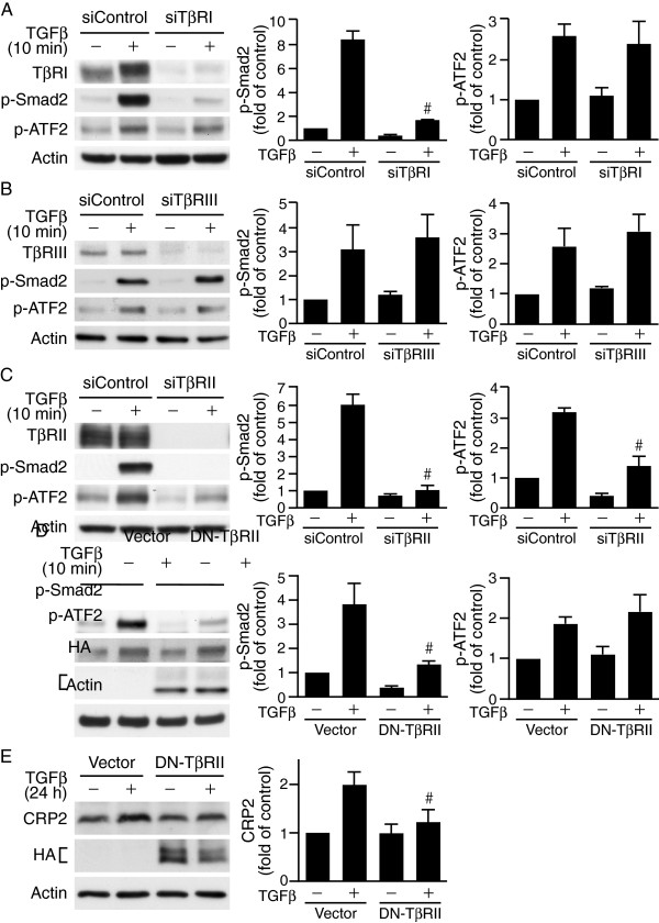
Figure 3.
Type II TGFβ receptor is crucial in mediating TGFβ-induced ATF2 signaling. (A-C) VSMCs were transfected with 20 nM control siRNA or siRNA to different types of TGFβ receptors. Following 24 h recovery in growth media and 24 h serum-starvation, cells were stimulated with or without TGFβ for 10 min. Western blot analysis was performed to detect different TGFβ receptor expression levels and phosphorylation of Smad2 and ATF2. (A) TβRI knockdown inhibits Smad2 phosphorylation but not that of ATF2. #P < 0.05 vs. TGFβ-stimulated siControl group. (B) TβRIII knockdown does not affect TGFβ activation of ATF2 or Smad2. (C) TβRII knockdown attenuates TGFβ activation of ATF2 and Smad2. #P < 0.05 vs. TGFβ-stimulated siControl group. (D) Dominant-negative TβRII (DN-TβRII) impairs Smad2 but not ATF2 activation. VSMCs were electroporated with control vector or HA-tagged DN-TβRII (HA-TβRII(∆Cyt)) and then stimulated with or without TGFβ for 10 min. Western blot analysis was performed to assess phosphorylation of Smad2 and ATF2. Overexpression of DN-TβRII was evaluated by probing Western blots with HA antibody. #P < 0.05 vs. TGFβ-stimulated vector control group. (E) DN-TβRII impairs TGFβ-induced CRP2 induction. VSMCs were electroporated with control vector or HA-TβRII(∆Cyt) and stimulated with TGFβ for 24 h. Total proteins were then isolated for Western blot analysis to detect CRP2 protein levels. The blots were then probed with HA antibody to detect HA-TβRII(∆Cyt) expression. #P < 0.05 vs. TGFβ-stimulated vector control group. The membranes were subsequently probed with actin for loading control. Representative blots of at least three independent experiments are shown.

