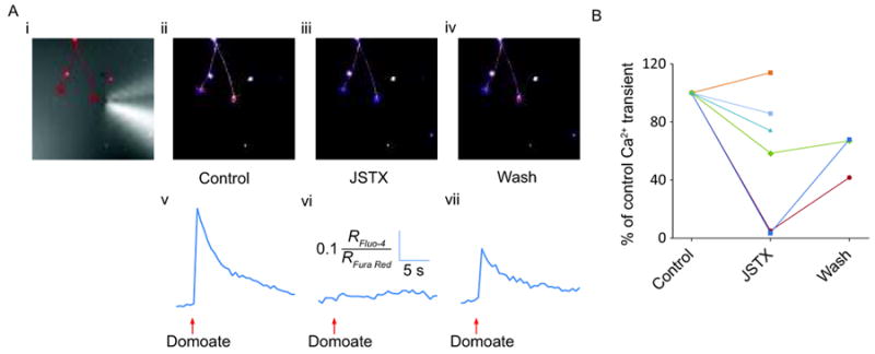
Figure 5.

Local domoate application and imaging at the growth cone in the absence and presence of JSTX. (i) DIC image of growth cone with an overlay of a thresholded fluorescence image showing the position of the puffer pipette tip close to the right-hand growth cone. Confocal images of the same cell obtained 1.2 s following domoate puff application in control (ii), after applying JSTX for 20 minutes (iii), and following a 20-minute wash after JSTX removal (iv). (v-vii) Corresponding traces showing fluorescence changes following domoate application in the conditions described in (i-iii). B) Graph showing the effect of JSTX on the amplitude of the domoate-induced fluorescence changes in 6 neurons. Fluorescence changes were normalized to the amplitude of fluorescence changes induced by the control application of domoate.
