
Fig. 7.
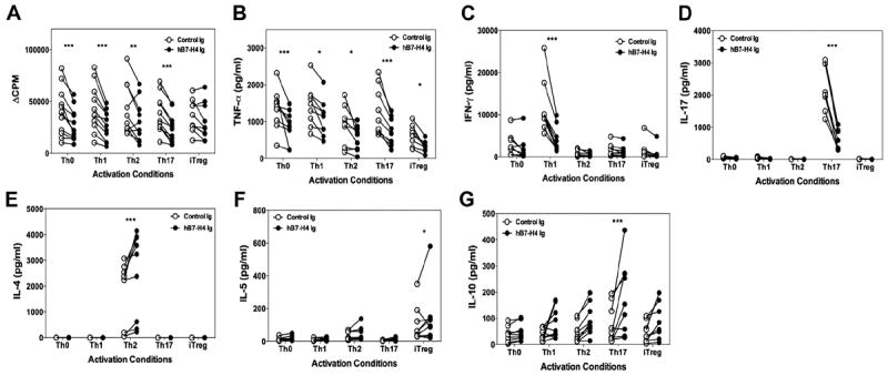
hB7-H4Ig decreases human Th1/Th17 cell differentiation and increases Th2 cell differentiation. Naive human CD4+ T-cells were isolated from total PMBCs collected from 10 to 12 healthy donors. The naive CD4+ T-cells (5 × 10⁵ cells/well) were activated in the presence of autologous irradiated total PBMCs (5 × 10⁵ cells/well) in the presence of anti-CD3 and anti-CD28 (0.5 μg/ml) plus Control-Ig or hB7-H4Ig (10 μg/ml). Cells were cultured in Th0- [IL-2 (100 U/ml), anti-IL-4, and anti-IFN-γ (1 μg/ml)], Th1- [IL-2 (100 U/ml), IL-12 (10 ng/ml), and anti-IL-4 (1 μg/ml)], Th2- [IL-2 (100 U/ml), IL-4 (10 ng/ml), and anti-IFN-γ (1 μg/ml)], Th17- [TGF-β (10 ng/ml), IL-6 (50 ng/ml), IL-1β (10 ng/ml), anti-IFN-γ, and anti-IL-4 (1 μg/ml)], or iTreg- [IL-2 (100 U/ml), TGF-β1 (25 ng/ml), and retinoic acid (100 nM)] promoting conditions. Two replicate sets of wells were set up such that one was (A) pulsed with 1 μCi of tritiated-thymidine at 24 h to determine cellular proliferation at 96 h, and for the second set the levels of (B) TNF-α, (C) IFN-γ, (D) IL-17, (E) IL-4, (F) IL-5, and (G) IL-10 were determined at 96 h post-culture initiation. Asterisks (*, **, ***) indicate a statistically significant alteration in the CPM values or cytokine level due to B7-H4Ig treatment as compared to Control-Ig treated cultures.