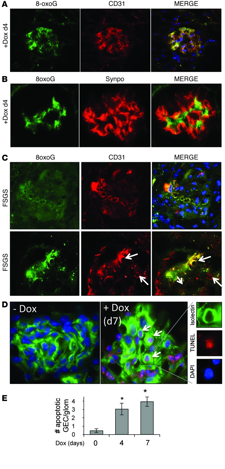
Figure 4. TGF-β signaling in podocytes induces oxidative stress specifically in endothelial cells.
(A) Representative double-immunofluorescence images of 8-oxoG (green), endothelial cell marker CD31 (red), and merge showing colocalization. (B) Representative double-immunofluorescence images of 8-oxoG (green), podocyte marker synaptopodin (red), and merge in glomeruli of a day 4 Dox-treated PodTgfbr1 mouse showing no colocalization in podocytes. (C) 8-oxoG (green) and endothelial cell marker CD31 (red) in glomeruli of kidney biopsies from subjects diagnosed with FSGS. Higher-magnification images showing capillary loops are shown on the bottom row (arrows) (original magnification, ×63). (D) Immunofluorescence staining showing TUNEL and isolectin localization with DAPI in untreated mice and mice treated with Dox for 7 days. Arrows depict nuclear TUNEL with DAPI- and isolectin-positive endothelial cells. (E) Quantification of TUNEL- and isolectin-positive cells per glomeruli in untreated PodTgfbr1 mice and in PodTgfbr1 mice treated with Dox for 4 or 7 days (*P < 0.05; mean ± SD). Original magnification, ×63 (A, B, C [bottom row], and D); ×40 (C [top row]).

