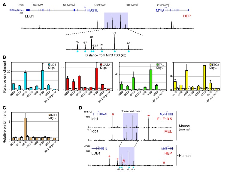
Figure 2. The HBS1L-MYB intergenic region contains regulatory elements bound by erythroid TFs.
(A) LDB1 ChIP-Seq data from primary HEPs. LDB1 peaks were marked by their distance to the MYB TSS. (B and C) ChIP-qPCR data (HEPs) showing enrichment (n = 3) for LDB1 complex members (B) and KLF1 (C) at the intergenic binding sites. IgG serum was used as control (IgG); the HBG1/2 promoter for normalization. (D) Comparison of mouse and human LDB1 ChIP-Seq data from erythroid progenitors. Binding sites not conserved are marked (*). The region containing the 4 conserved sites (conserved core) is highlighted in purple. Error bars display SEM. FL E13.5, 13.5 dpc fetal liver erythroid progenitors.

