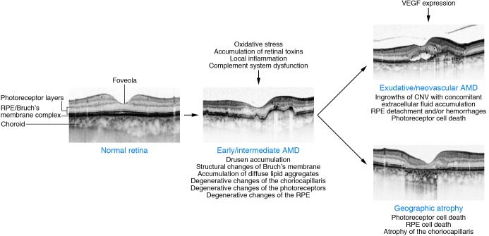
Figure 1. Schematic showing morphological changes in the macula during evolution of early/intermediate AMD, exudative/neovascular AMD, and GA, respectively, along with several known pathogenetic factors.
The course of the disease, however, can vary from case to case. High-resolution SD-OCT images show typical findings of the different AMD stages. Images reproduced with permission from Investigative Ophthalmology Visual Science (85).

