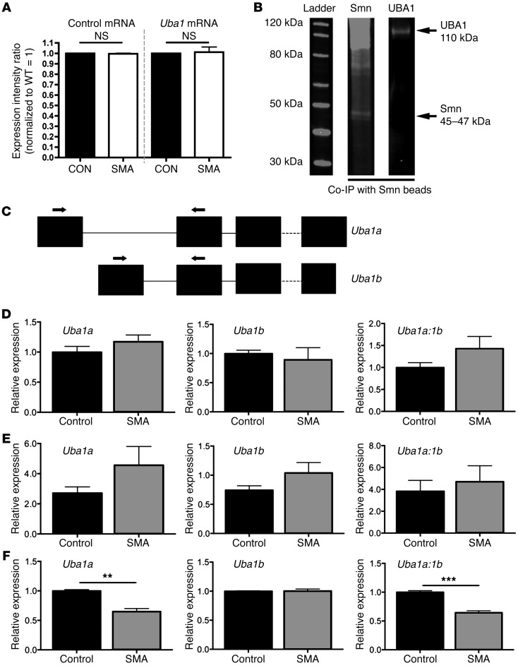
Figure 2. UBA1 physically interacts with SMN protein in vivo, and Uba1 splicing is dysregulated at late symptomatic time points in SMA mouse spinal cord.
(A) No change in levels of Uba1 mRNA (or a control mRNA, Fth1; similar control data using Mapt not shown) in the spinal cord of P5 severe SMA mice, quantified using qPCR (n = 3 mice per genotype; ANOVA with Tukey’s post hoc test). (B) Representative fluorescent Western blots for SMN (left lane) and UBA1 (right lane) from co-IP experiments on spinal cord extracts from WT mice, using SMN-bound beads, demonstrating that UBA1 physically interacts with SMN in vivo. (C) Graphic overview of the exon structure of Uba1. Two Uba1 splice variants are generated with unique first exons. The position of primers used to amplify each splice variant is highlighted. Note that the coding sequence of Uba1 starts in exon 2. (D–F) Bar charts showing relative expression levels of Uba1a and Uba1b, as well as the ratio of Uba1a to Uba1b, in SMA (Taiwanese) and control spinal cord at P3 (D; presymptomatic), P7 (E; early symptomatic), and P11 (late-symptomatic) (n = 3 mice per genotype, 3 independent amplifications per sample; 2-tailed, unpaired t tests). Uba1 splicing was significantly dysregulated in the late-symptomatic mice. **P < 0.01; ***P < 0.001.

