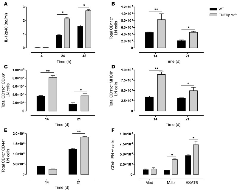
Figure 5. Increased activated DCs induces enhanced M. tuberculosis–specific IFN-γ production by activated CD4+ T cells in M. tuberculosis–infected TNFRp75–/– mice.
(A) BMDCs from WT and TNFRp75–/– mice were infected at an MOI of 5:1 with M. tuberculosis, and IL-12p40 expression in culture supernatants was analyzed by ELISA. (B–E) WT and TNFRp75–/– mice were infected at 50–100 CFU with M. tuberculosis, and lungs and LNs were harvested 14 and 21 days after infection. The total number of LN CD11c+ cells (B), CD11c+CD86+ cells (C), CD11c+MHCII+ cells (D), and CD4+CD44+ T cells (E) was analyzed by flow cytometry. Data (mean ± SEM of 4 mice per group) are representative of 3 similar experiments. (F) IFN-γ expression by CD4+ T cells in isolated pulmonary cell cultures restimulated with ESAT6 or M. tuberculosis H37Rv (M.tb). Data (mean ± SEM of triplicate values of pooled samples from 4 mice) are representative of 3 similar experiments. *P < 0.05, **P < 0.01, ANOVA.

