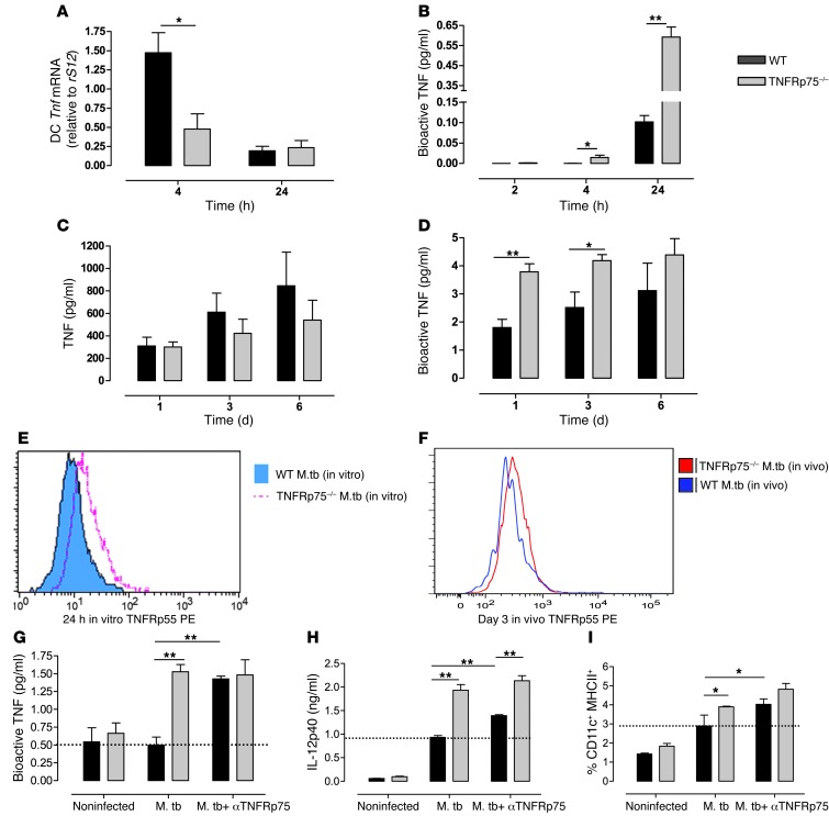
Figure 7. Soluble TNFRp75 inhibits DC activation during M. tuberculosis infection.
(A and B) WT and TNFRp75–/– DCs were infected with M. tuberculosis at an MOI of 5:1, and Tnf mRNA (A) or bioactive TNF (B) concentrations were measured. (C and D) WT and TNFRp75–/– mice were infected with 50–100 CFU M. tuberculosis. Total (C) and bioactive (D) TNF was measured in BALF 1, 3, and 6 days after infection. (E and F) In vitro surface expression of TNFRp55 (E) was measured in WT and TNFRp75–/– BMDCs, and in vivo surface TNFRp55 (F) was measured by flow cytometry in BAL cells. (G–I) The effect of soluble TNFRp75 on M. tuberculosis–infected BMDC cultures from WT and TNFRp75–/– mice were assessed by measuring bioactive TNF (G), IL-12p40 (H), and MHCII+ expression (I) in the presence or absence of anti-TNFRp75. Note the increased expression induced by anti-TNFRp75 in WT mice compared with M. tuberculosis infection alone (dashed lines). Data (mean ± SEM of quadruplicate experiments) are representative of 1 of 2 similar experiments. *P < 0.05, **P < 0.01, ANOVA.

