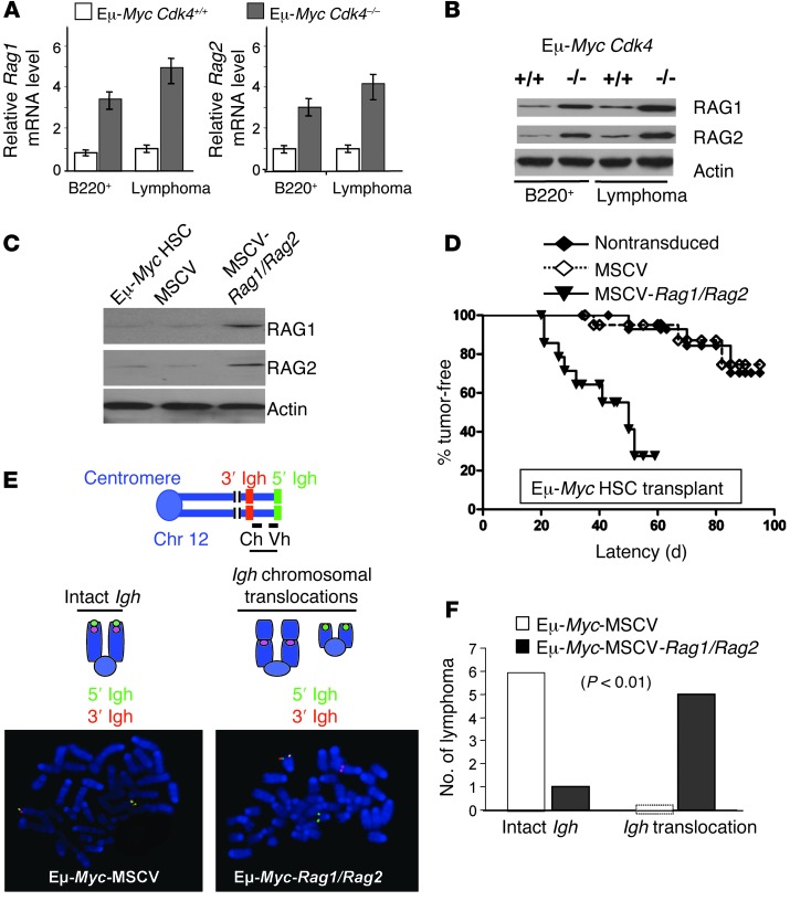
Figure 4. CDK4 controls RAG1 and RAG2 expression in Eμ-Myc B cells, and forced coexpression of RAG1 and RAG2 is sufficient to augment the development of Myc-driven lymphoma.
(A) Rag1 and Rag2 mRNA levels in Eμ-MycCdk4–/– and Eμ-MycCdk4+/+ premalignant bone marrow B220+ and lymphoma cells (n = 5 mean ± SD, P < 0.005, respectively). (B) RAG1 and RAG2 protein levels in Eμ-MycCdk4–/– and Eμ-MycCdk4+/+ premalignant B220+ and lymphoma cells (data shown are representative of analyses of five cohorts of Eμ-MycCdk4–/– and Eμ-MycCdk4+/+ B220+ B cells and lymphomas). (C) Overexpression of RAG1 and RAG2 in Eμ-Myc HSCs. HSCs from E13.5–E15.5 Eμ-Myc fetal livers were transduced with MSCV-IRES-Puro (MSCV) control virus or were cotransduced with MSCV-Rag1-IRES-Puro and MSCV-Rag2-IRES-Hygro (MSCV-RAG1/RAG2) retroviruses. Lysates from these HSCs were analyzed by Western blotting. (D) Enforced coexpression of RAG1 and RAG2 accelerates lymphoma development. 3 × 10⁶ HSCs (nontransduced, MSCV, or MSCV-Rag1/Rag2-HSCs; n = 15) were transplanted into lethally irradiated congenic recipients that were then followed daily for lymphoma onset (P < 0.001). (E) FISH analyses of lymphomas arising in recipient mice engrafted with Eμ-Myc HSCs transduced with MSCV control retrovirus or cotransduced with MSCV-RAG1/RAG2-expressing retroviruses. Metaphase cells from each cohort were assessed by FISH with 5′ (green) and 3′ (red) Igh probes. An intact Igh locus had colocalized red and green signals, while a broken, translocated locus had split red and green signals. Original magnification, x1,500. (F) Statistical analysis of Igh translocations in the two cohorts of lymphomas, Eμ-Myc-MSCV and Eμ-Myc-MSCV-RAG1/RAG2 (n = 6, P < 0.01).

