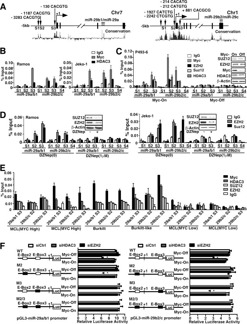
Figure 3. Myc recruits HDAC3 and PRC2 to miR-29 promoters to repress the miR-29 transcription through histone deacetylation and trimethylation.
(A) Schematic diagram showing location of Myc-binding sites of pri-miR-29a/b1 and pri-miR-29b2/c regulatory region. S1, S2, and S3 represent Myc-binding site, which has E-box sequence. S4 was used as negative control and is located in the intron 4 of pri-miR-29b2/c and without E-box in this region. Both pri-miR-29s are highly conserved in their putative promoter region and in the pre-miR-29 stem sequences, encoded in the last intron (pre-miR-29a/b1) on chr.7q32.3 and the last exon (pre-miR-29b2/c) on chr.1q32.2 respectively. (B) ChIP assay showing Myc and HDAC3 enrichment on pri-miR-29a/b1 and pri-miR-29b2/c promoters. ChIP assay was performed using Myc or HDAC3 antibody to detect binding on pri-miR-29a/b1 and pri-miR-29b2/c promoters, S1-S3 regions and S4 was used as a negative control. % Input was calculated with 2(Ct [1% of input]-Ct[ChIP]). (C) ChIP assay showing Myc, HDAC3, EZH2 and SUZ12 enrichment on pri-miR-29a/b1 and pri-miR-29b2/c promoters and dependence of HDAC3, EZH2/SUZ12 binding on Myc in P493-6 cells with or without 24 hours Tet treatment, Inserts, Western blots showing protein level of Myc, HDAC3 and EZH2/SUZ12 in Myc-on and Myc-off (24 hours Tet treatment) P493-6 cells. (D) ChIP assay showing EZH2 and SUZ12 enrichment on pri-miR-29a/b1 and pri-miR-29b2/c with or without DZNep treatment. (E) ChIP assay showing Myc, HDAC3, EZH2 and SUZ12 enrichment on pri-miR-29a/b1 and pri-miR-29b2/c promoters in primary lymphoma samples with high Myc expression (blastic MCLs, Burkitt or Burkitt like lymphomas) and no enrichment in primary samples with low myc expression (indolent MCLs). (F) Schematic diagram of pri-miR-29a/b1 and pri-miR-29b2/c promoter luciferase reporter. Solid boxes represent point mutation of E-Box. P493-6 cells were transfected with either wild-type or mutants (M) of pri-miR-29a/b1 or pri-miR-29b2/c promoter luciferase reporter, together with siHDAC3, siEZH2, or non-targeting siRNA. The luciferase activity is normalized to β-galactosidase. Results are means ± SD from 3 biological replicates. For ChIP assays, IgG was used as negative control. In B-F, results are means ± SD from at least 3 biological replicates. Inserts, Western blots showing protein level. (See also Figure S3)

