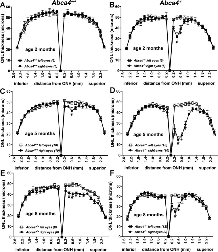
Figure 2.
ONL thicknesses measured in Abca4+/+ (A, C, E) and Abca4−/− (B, D, F) mice, both light-stressed (right) and control (non–light-stressed; left) eyes of 2-, 5-, and 8-month-old mice 7 days after light exposure. Values are mean ± SEM of numbers of eyes presented in parentheses. *P < 0.05 when right eyes were compared to left eyes by one-way ANOVA and Sidak's multiple comparison test. The lesions present as areas of reduced ONL thickness in superior retinae.

