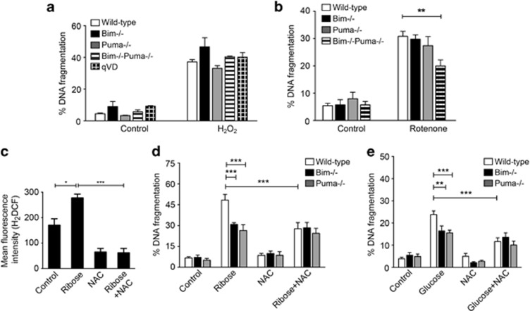
Figure 6.
Inhibition of oxidative stress protects islet cells from glucotoxicity. (a and b) The frequency of cells undergoing DNA fragmentation was measured by flow cytometry after incubation of wild-type C57BL/6, Bim−/−Puma−/−, Bim−/− or Puma−/− islets with 30 μM H2O2 and 50 μM pancaspase inhibitor qVD.oph for 2 days (a), or 100 nM Rotenone for 2 days (b). Control islets were incubated for 2 days in a medium containing 5.5 mM glucose. Results represent mean±s.e.m. of n≥4 independent experiments. **P<0.01 ***P<0.001 (two-way ANOVA). (c) Mean fluorescence intensity of H2DCF staining measured by flow cytometry after incubation of MIN6 cells with 50 mM ribose with or without 1.0 mM NAC for 3 days. Results are mean±s.e.m. of n=3–4 independent experiments. *P<0.05, ***P<0.001 (one-way ANOVA). (d and e) The frequency of cells undergoing DNA fragmentation was measured by flow cytometry after incubation of wild-type C57BL/6, Puma−/− or Bim−/− islets with 50 mM ribose with or without 1.0 mM NAC for 4 days (d), or 33.3 mM glucose with our without 1.0 mM NAC for 6–7 days (e). Control islets were incubated in a medium containing 5.5 mM glucose. Results represent mean±s.e.m. of n≥5 independent experiments. **P<0.01, ***P<0.001 (two-way ANOVA)

