Figure 6.
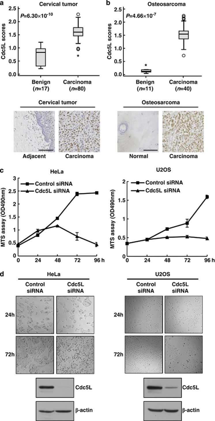
Cdc5L is overexpressed in cervical tumors and osteosarcoma and its depletion decreases cell viability of related tumor cells. (a) The tissue array of cervical tumor was performed by immunohistochemistry with anti-Cdc5L antibody. Cdc5L expression was plotted using the score as described in the ‘Materials and Methods' section. Outliers are indicated by open circles, extremes by asterisks (top panel). Representative images from immunohistochemical staining of Cdc5L in adjacent tissue and cervical tumor; scale bar, 100 μm (bottom panel). (b) The tissue array of osteosarcoma was performed as in a. Cdc5L expression was plotted using the score as described in the ‘Materials and Methods' section. Outliers are indicated by open circles, extremes by asterisks (top panel). Representative images from immunohistochemical staining of Cdc5L in normal bone and osteosarcoma; scale bar, 50 μm (bottom panel). (c) Cell viability assay in control or Cdc5L-knockdown HeLa or U2OS cells was measured by MTS at indicated times after siRNA transfection. Data are shown as mean±S.D. (d) Representative images of cell growth inhibition in control and Cdc5L-knockdown cells at indicated times (top panel); immunoblot analysis of knockdown efficiency of Cdc5L in indicated cells (bottom panel)

