Figure 6.
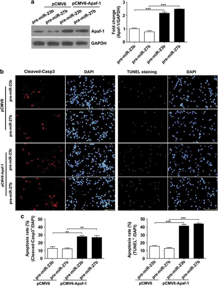
Overexpression of Apaf-1 re-activates the sensitivity of neurons overexpressing miR-23b/27b to apoptosis induced by hypoxia. (a) Western blot analysis of Apaf-1 in primary cortical neurons 48 h after co-transfection with plasmids (pCMV6 or pCMV6-Apaf-1) and miRNAs (pre-miR-23b or pre-miR-27b). Relative amounts of Apaf-1 protein were quantified by densitometry (n=3, unpaired t-test, ***P<0.001). (b) Immunofluorescence staining of Cleaved-Casp3 or TUNEL staining. Cultured cortical neurons co-transfected with plasmids (pCMV6 or pCMV6-Apaf-1) and miRNAs (pre-miR-23b or pre-miR-27b) were stained with Cleaved-Casp3 antibody or with TUNEL staining kit after hypoxia treatment (24 h) at DIV5. (c) Statistical analysis of the percentage of Cleaved-Casp3-positive neurons or TUNEL-positive neurons to DAPI-stained cells (cell numbers=700–800, unpaired t-test, **P<0.01 and ***P<0.001). Scale bar represents 100 μm

