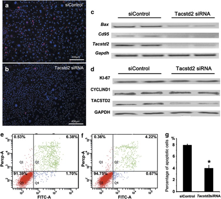
Figure 6.
Effects of TACSTD2 on apoptosis and proliferation. (a and b) Immunofluorescent staining of cleaved caspase-3 (red) in control and Tacstd2-silenced HaCaT cells. Nuclei were counterstained with DAPI (blue). Two Tacstd2 siRNA oligos were tested as described in Materials and Methods section, and similar phenotype were observed. Higher efficiency of Tacstd2 knockdown was observed by using Tacstd2 siRNA oligo (ID HSS106222). (c) RT-PCR analysis of Bax, Cd95 and Gapdh mRNAs in control and Tacstd2-silenced HaCaT cells. (d) Western blot analysis of KI67, CYCLIND1, TACSTD2 and GAPDH in control and Tacstd2-silenced HaCaT cells. (e and f) Representative figures for gemcitabine-induced apoptosis, analyzed by flow cytometry using Annexin V-FITC/PI, are shown for control and Tacstd2-silenced HaCaT cells. (Q1: necrosis; Q2: late apoptosis; Q3: healthy cells; Q4: early apoptosis). (g) A bar graph showing percentage of gemcitabine-induced apoptosis in control and Tacstd2-silenced HaCaT cells. A significant difference was identified between control and Tacstd2-silenced HaCaT cells by using the Student's t-test (*P<0.05, N=3)

