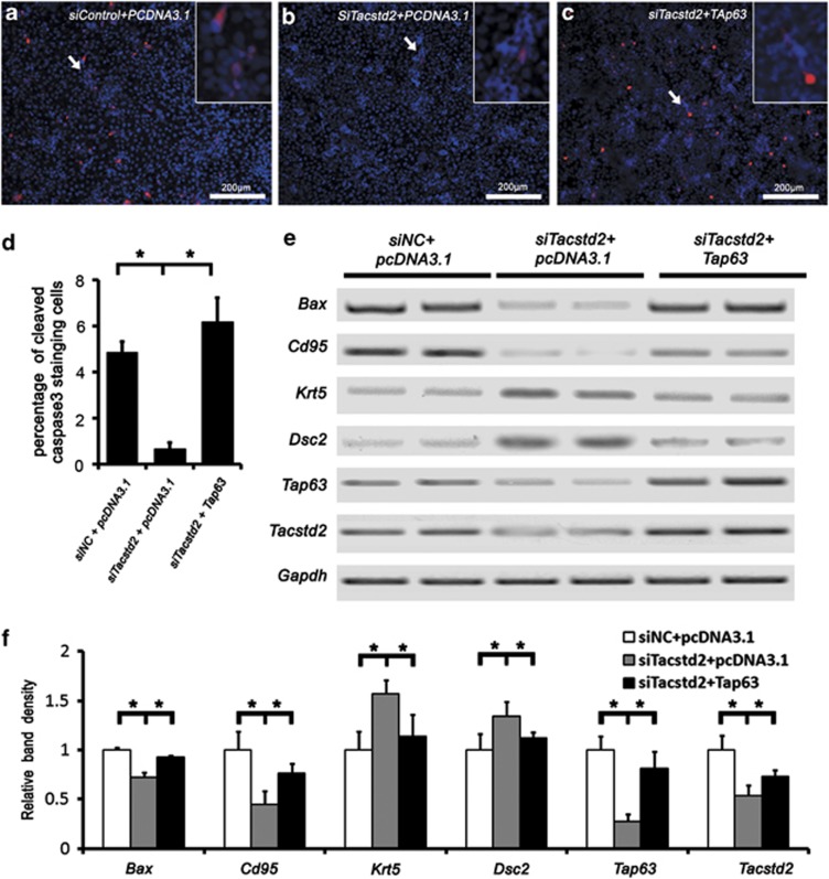
Figure 7.
Induced expression of TAp63 in HaCaT cells rescued TACSTD2-dependent caspase-3 activation and apoptotic gene expression. (a–c) Immunofluorescent staining of cleaved caspase-3 (red). Nuclei were counterstained with DAPI (blue). Inserts are higher magnifications of the cleaved caspase-3-positive cells. (d) A bar graph showing percentage of cleaved caspase-3-positive cells. Significant differences were identified between control and Tacstd2-silenced HaCaT cells, between Tacstd2-silenced HaCaT cells and TAp63 rescued cells by using the Student's t-test (*P<0.05, N=3). (e) RT-PCR analysis of Bax, Cd95, TAp63, Krt5, Dsc2, Tacstd2 and Gapdh. (f) A bar graph showing relative mRNA levels. Significant differences were identified between control and Tacstd2-silenced HaCaT cells, between Tacstd2-silenced HaCaT cells and TAp63 rescued cells by using the Student's t-test (*P<0.05, N=3)

