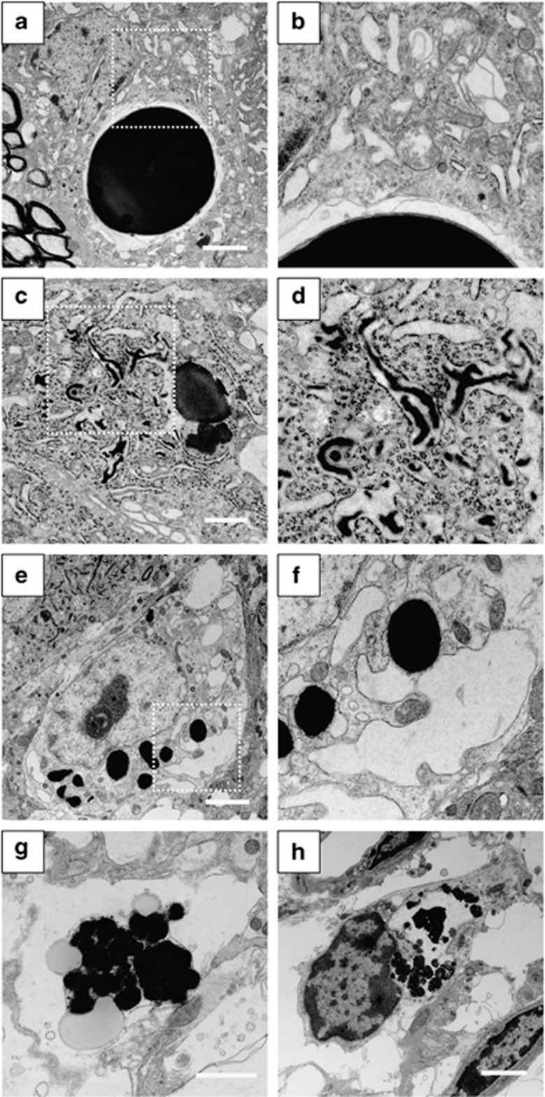
Figure 7.
Electron microscopic analysis in AVP neurons of 5-month-old FNDI mice. AVP neurons of the SON in 5-month-old FNDI mice with water access ad libitum (a–d) and of those subjected to WD for 12 weeks (e–h). Higher magnification images of boxed areas in (a, c and e) are shown in (b, d and f), respectively. Scale bars, 2 μm (a, e, g and h), 1 μm (c)

