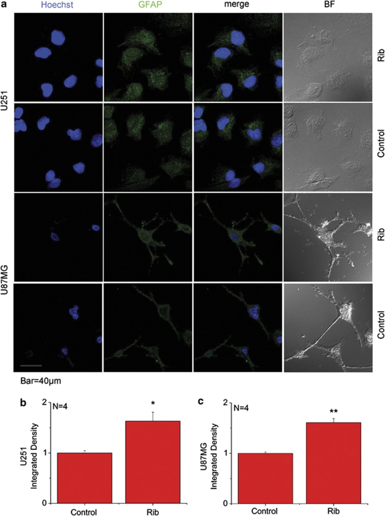
Figure 3.
GFAP immunofluorescence staining on Rib-treated astrocytoma cells. U251 and U87MG cells (a) were treated with 20 mM Rib for 2 days. Untreated cells were used as controls. The distribution of GFAP was detected by immunofluorescent staining using an anti-GFAP antibody. Cell nuclei were stained with the DNA-specific fluorescent reagent Hoechst 33258. Quantification results were shown in b and c, respectively. The control value was set as 1.0. All values are expressed as means±S.E.M. *P<0.05, **P<0.01

