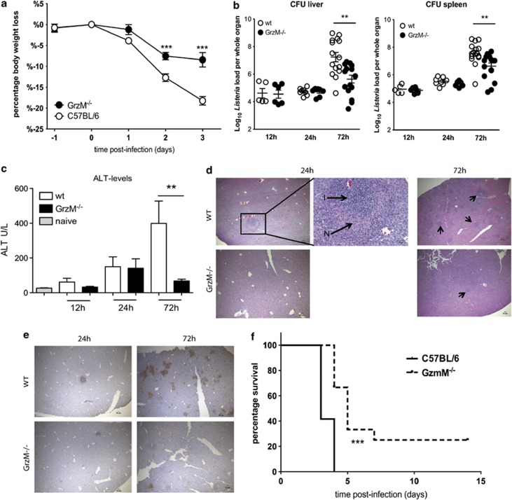
Figure 1.
GrzM−/− mice are less susceptible to an infection with L. monocytogenes. (a–e) Female, age-matched WT and GrzM−/− mice were infected i.v. with 104 CFU of L. monocytogenes. (a) The recipients were monitored daily for weight loss and signs of illness. (b) After 12 h, on days 1 and 3 after infection mice were killed, livers and spleens were homogenized, serially diluted in PBS and plated on 5% sheep agar plates. Bacterial colonies were counted after 24 h of incubation at 37 °C. (c) Blood samples were taken 12, 24 and 72 h after infection and ALT levels were determined using the Architect plus analyser. (d) H & E staining of liver sections of WT and GrzM−/− mice 24 and 72 h after Listeria infection (N=necrotic foci, I=inflammatory infiltrations) (representative). Arrows indicate inflammatory foci. (e) TUNEL+ staining of WT and GrzM−/− mice 24 and 72 h after Listeria infection (representative). Pictures were taken with an Olympus BX-51, original magnification for images × 4 (NA 0.13, WD17 mm) or × 20 (NA 0.7, WD 0.65 mm). (f) The survival of recipients was monitored. Mice were killed when they had lost 20% of their initial body weight. Results are pooled (a) from two independent experiments with n=10, (b and c) from six independent experiments with n=5–15, (f) from two independent experiments, n=12

