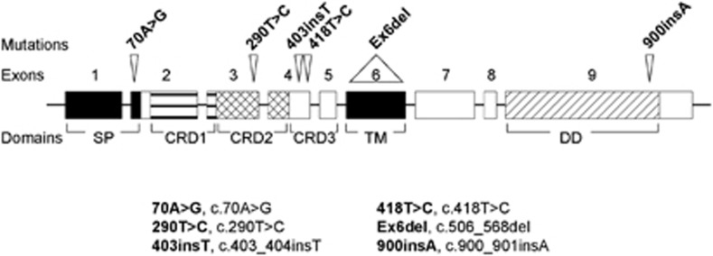
Figure 1.

FAS mutations in T-LBL samples. Schematic representation of mutations found in FAS in human T-LBL samples, indicating FAS exons and FAS protein domains affected. An abbreviated form to indicate the mutations is included and explained in the legend
