Figure 4.
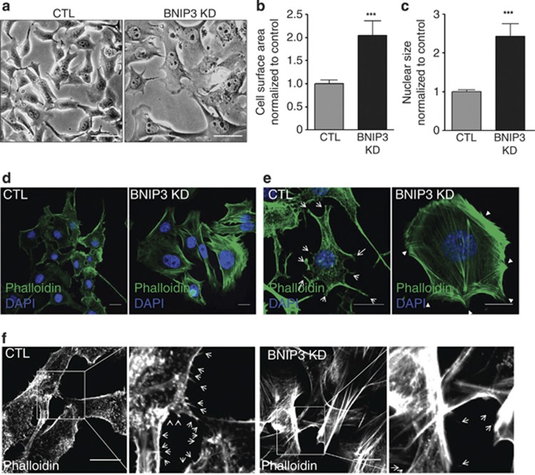
BNIP3 is required to maintain cellular architecture and cytoskeletal structures. (a) Representative light microscopy images (n=4, five pictures per experiment) of control and BNIP3 KD B16-F10 cells (24 h after cell plating), scale bars represent 50 μm. (b) Cell surface area, quantified using ImageJ, of control and BNIP3 KD cells. (c) Nuclear size, quantified using Image J, of control and BNIP3 KD cells. (d) Confocal microscopy analysis of the actin cytoskeleton of B16-F10 cells (24 h after cell plating) using the high affinity actin-probe phalloidin-Alexa Flour 488 and the nuclear dye DAPI. Representative images (n=5, five pictures per condition) of control versus BNIP3 KD cells, scale bars represent 10 μm. (e) Confocal microscopy analysis of the actin cytoskeleton using the high affinity actin-probe Phalloidin-Alexa Flour 488 and the nuclear dye DAPI on low density cell culture of B16-F10 cells (24 h after cell plating). Representative images (n=5, five pictures per condition) of control versus BNIP3 KD cells are shown. Scale bars represent 10 μm. Arrows indicate lamellipodia and arrowheads indicate membrane ruffles. (f) Higher magnification images of the staining in e to reveal cell filopodia. Representative images (n=5, five pictures per condition) and zoom of control and BNIP3 KD B16-F10 cells are shown, scale bar represents 10 μm, arrows indicate filopodia

