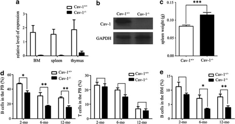
Figure 1.
The percentage of B cells from the peripheral blood, BM and spleen was decreased in Cav-1−/− mice. (a) Expression of the Cav-1 protein in the BM, spleen and thymus was detected using real-time PCR. (b) Expression of the Cav-1 protein in the BM was detected using western blotting. (c) The weight of the spleen from Cav-1+/+ and Cav-1−/− mice. (d) FACS analysis of the percentage of B and T cells in the peripheral blood from 2-mo-, 6-mo- and 12-mo-old mice. (e) FACS analysis of the percentage of B cells in the BM from 2-mo-, 6-mo- and 12-mo-old mice; n=5 mice per group. The experiment was repeated three times, and the results represent the mean±s.d. *P<0.05; **P<0.01; ***P<0.001. GAPDH expression was used for normalization

