Figure 2.
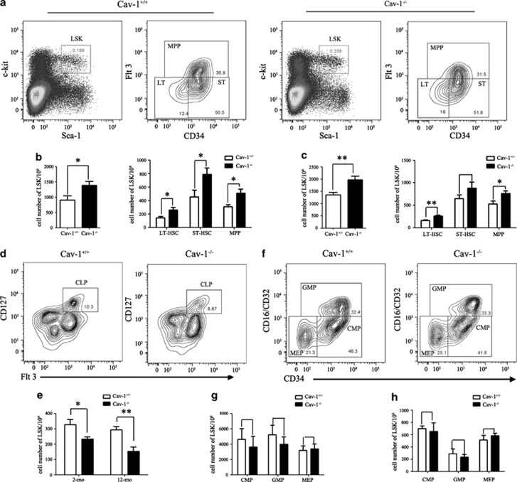
Hematopoietic stem/progenitor cell defects in Cav-1−/− mice. (a) Representative staining profiles for BM HSCs and progenitor populations. (b) Cell numbers of LSKs, LT-HSCs, ST-HSCs and MPPs in the BM from 2-mo- and 12-mo-old (c) mice. (d) Representative staining profiles for CLPs (Lin−Sca-1lowc-KitlowCD127+) in the BM. (e) Cell numbers of CLPs in the BM from 2-mo- and 12-mo-old mice. (f) Representative staining profiles for myeloid progenitors. L−S−K+ cells were subdivided into CMPs (Lin−Sca-1−c-Kit−CD34+CD16/CD32−), GMPs (Lin−Sca-1−c-Kit−CD34+CD16/CD32+) and MEPs (Lin−Sca-1−c-Kit−CD34−CD16/CD32−) as indicated. (g) Cell numbers of CMPs, GMPs and MEPs in the BM from 2-mo- and 12-mo-old (h) mice. n=5 mice per group. The experiment was repeated three times, and the results represent the mean±s.d. *P<0.05; **P<0.01

