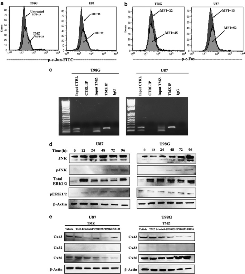
Figure 4.
EGFR-mediated activation of AP-1 in TMZ-treated GBM cells. (b) U87 and T98G cells were treated with 200 μM TMZ. After 72 h, the viable cells were analyzed by flow cytometry for phospho (p)-c-Jun (a) and p-c-Fos activation. The dark histograms represent untreated cells and the gray histograms, TMZ treatment. The MFI of each histogram is depicted with an arrow. (c) ChIP analyses were performed for AP-1 binding to endogenous Cx43 using TMZ-resistant GBM cells. The complex was precipitated with anti-c-Jun. Shown are the PCR of the precipitated gDNA with primers spanning the AP-1 site. (d) Western blots were performed for the upstream activators of AP-1 activation, pERK and p-JNK using whole-cell extracts from untreated (Time 0) and timeline treatment of U87 and T98G with 200 μM TMZ. The timeline studies occurred at 12 h intervals up to 96 h. (e) GBM cells were pretreated different pharmacological agents along the EGFR signaling pathway. After 24 h, the cells were washed and then exposed to 200 μM TMZ for 72 h. Whole-cell extracts were analyzed by western blots for Cx26, Cx32 and Cx43

