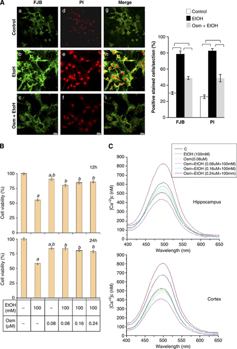
Figure 1.
Osmotin protects against ethanol toxicity to primary cultures of fetal neurons. (A) Fluorescence analysis of neurodegeneration in primary cultures of fetal rat brain hippocampal neurons. Cultures were exposed to growth medium without (Control) and with ethanol (EtOH, 100 mM) or osmotin (Osm, 0.16 μM) plus EtOH (100 mM) supplements for 24 h before staining with Fluoro-Jade B (FJB; green) and propidium iodide (PI; red). Magnification × 60; scale bar, 20 μm. Shown in the graph are the percentages of FJB- and PI-positive cells per section (n=4). Indicated pairs are significantly different at P<0.05. (B) MTT assay of cell viability in primary cultures of fetal rat brain hippocampal neurons. Cell viability was measured following exposure to ethanol and osmotin at the indicated concentrations for 12 and 24 h. Data are the mean±S.E. of three independent experiments (n=3), with three plates in each experiment. Statistically significantly differences at P<0.05 are indicated by symbols. Symbols: a, different from control; b, different from ethanol (100 mM). (C) Depicted are the analysis of cytosolic calcium concentrations in primary cultures of fetal rat brain hippocampal and cortical neurons exposed to the indicated treatments for 24 h, followed by fura-2 AM labeling

