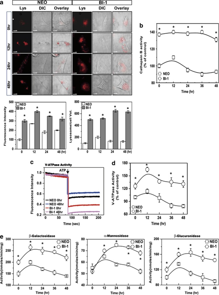
Figure 3.
BI-1 is associated with high lysosomal activity. (a) Neo and BI-1 cells were cultured in serum-free medium with or without 5 ng/ml TGF-β1 for 0, 12, 24, or 48 h. Then, cells were exposed to 5 μM LysoTracker and photographed. Scale bar, 30 μm. Fluorescence intensity and volume were quantified (bottom). *P<0.05, significantly different from Neo cells at each point. (b) After isolation of lysozymes, cathepsin B was analyzed. For lysosomal V-ATPase activity, 6 μM acridine orange solution was added to lysosomal membranes from Neo and BI-1 cells that were untreated or treated with TGF-β1 for 48 h. (c) Intra-vesicular H+ uptake was initiated by the addition of Mg-ATP, and fluorescence was measured as described in the Materials and Methods. (d) V-ATPase activity was quantified in TGF-β1-treated and non-treated Neo and BI-1 cells for the indicated periods. (e) β-Galactosidase, α-mannosidase, and β-glucuronidase activity in the lysosomal extracts was measured using a spectrofluorospectrometer. Lys, LysoTracker

