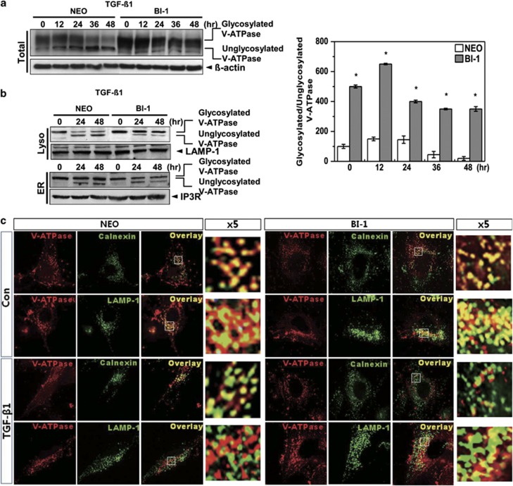
Figure 5.
Lysosomal V-ATPase is highly expressed in BI-1 cells. (a) Neo and BI-1 cells were cultured in serum-free medium with or without 5 ng/ml TGF-β1 for 0, 12, 24, 36, or 48 h. Immunoblotting was performed with antibodies against the V-ATPase V0a1 subunit or β-actin. Ratio of glycosylated to unglycosylated V-ATPase was quantified as shown in the right panel. *P<0.05, significantly different from Neo cells during each period. (b) Neo and BI-1 cells were cultured in serum-free medium with or without 5 ng/ml TGF-β1 for 0, 24, or 48 h. After the lysosome and ER fractions were isolated, immunoblotting was performed using anti-V-ATPase V0a1 subunits, LAMP-1, or IP3R antibodies. (c) Neo and BI-1 cells were cultured in serum-free medium with or without 5 ng/ml TGF-β1 for 48 h. Immunostaining with anti-V-ATPase, calnexin, or LAMP-1 antibodies was performed, and the images were captured under a fluorescent microscope

