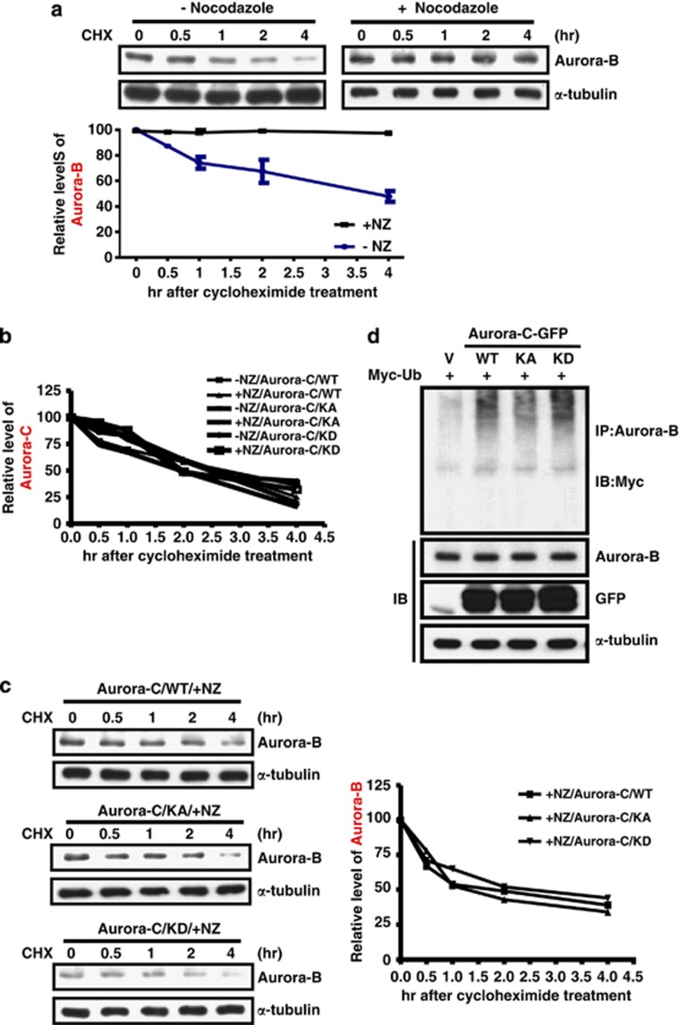
Figure 6.
Aurora-B protein stability is enhanced upon SAC activation and is reduced in cells overexpressing Aurora-C. (a) HeLa cells with or without nocodazole treatment were further incubated with cyclohexamide (CHX) at different time points. Total lysates were prepared for IB analysis using anti-Aurora-B. The expression levels of Aurora-B were normalized by α-tubulin. The quantitative results of three independent experiments are shown below. (b) Cells expressing Aurora-C/WT, KA, or KD were treated as described above. Aurora-C expression levels were determined using IB analysis and normalized by α-tubulin. Quantitative results are shown. (c) The expression levels of Aurora-B in cells expressing Aurora-C/WT, KA, or KD expression were determined by IB analysis and normalized by α-tubulin. All cells were treated with nocodazole, followed by CHX as described above. Quantitative results are shown. (d) Cells were co-transfected with Aurora-C-GFP/WT, KA or KD, and Myc-Ubi. Equal amounts of total lysates were used for the IP assay using anti-Aurora-B antibody followed by IB analysis using anti-Myc antibody. The expressions of Aurora-B, Aurora-C-GFP/WT, KA, KD, and α-tubulin were determined using IB analysis

